Anatomy of the spine
The spine is made of 33 individual bones one above the other. This spinal column provides support for the entire body allowing to stand upright. It also helps in bending, and twisting. It protects the spinal cord from injury. An adult spine has a natural S-shaped curve. The curves helps to absorb shock, maintain balance, and allow the range of motion throughout the spinal column.
Vertebrae
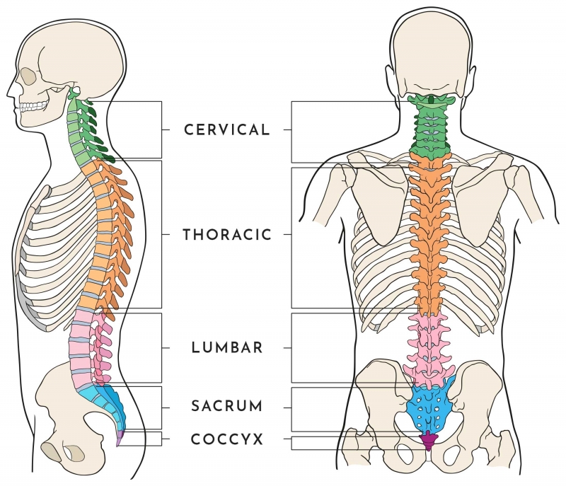
Vertebrae are the 33 individual bones that interlock with each other to form the spinal column.
- Cervical – 07 bones – They supports the head and helps in movements of neck
- Thoracic – 12 bones – They hold the rib cage and protect the heart and lungs
- Lumbar – 05 bones – They bear the weight of the body and helps in bending
- Sacrum – 05 bones – They connect the spine to the hip and form pelvic girdle
- Coccyx – 04 bones – they form tail bone and aid in ligament attachments
Intervertebral discs
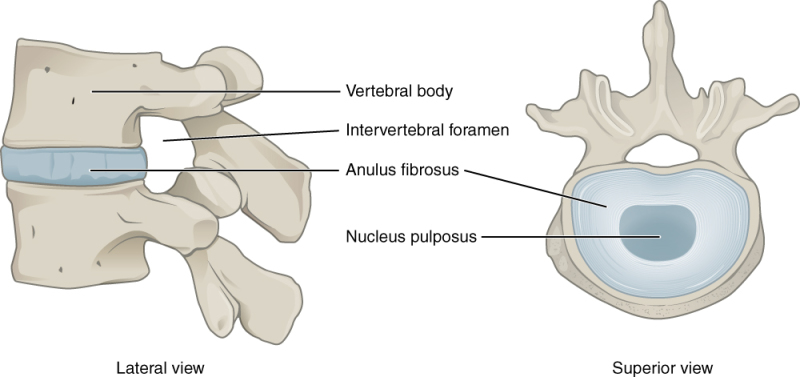
Intervertebral disc separates the adjacent two vertebral bones. Discs have two pars (i) The outer ring, called the annulus (ii) inner gel-filled centre called the nucleus.
Discs function like springs or cushions. With age, our discs increasingly become rigid and lose elastic properties. Injury and strain to discs can cause them to bulge/herniate through the annulus to compress the nerve roots causing back pain.
Spinal canal
The hollow spinal canal in the vertebral bone contains the spinal cord, fat, ligaments, and blood vessels. Below the pedicle, spinal nerves exits the spinal cord and pass through the intervertebral foramen to respective part of the body.
Facet joints & ligaments
The facet joints present on either side of the spine allow back motion. The ligamentum flavum, anterior longitudinal ligament and posterior longitudinal ligament are strong fibrous bands that hold the vertebrae together, stabilize the spine, and protect the discs.
Spinal cord
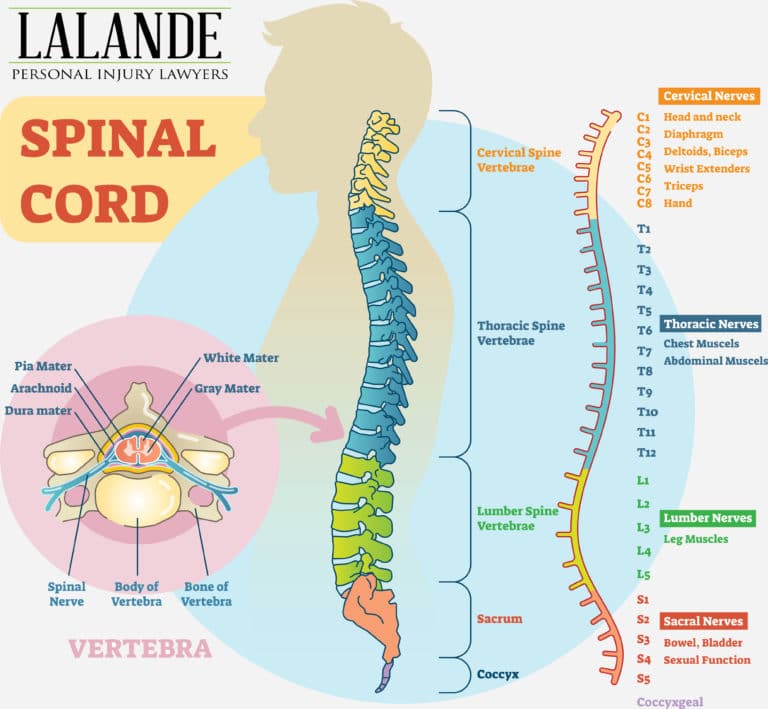
The spinal cord runs from the brainstem to the first lumbar vertebra within the spinal canal. At the end of the spinal cord, the nerve fibers fill the spinal canal, which are called cauda equine. They continue down through the spinal canal to your tailbone before branching off to your legs and feet. The spinal cord send impulses of sensation from the body to the brain. The brain sends motor impulses to the limbs and body through the spinal cord.
Spinal nerves
A each vertebrae, one pair of spinal nerves branch out, a total of Thirty-one pairs of spinal nerves.The nerve passes through the intervertebral foramen, it branches; each branch has both motor and sensory fibers. The spinal nerves innervate specific areas and form a striped pattern across the body called dermatomes. This pattern helps to diagnose the location of a spinal problem based on the area of pain or muscle weakness.
GENERAL ENQURIES
186 0208 0208
+91 8884415615 | +91 8884458890
- gouthamcugati@gmail.com
- Narayana Multispecialty Hospital.
CAH/1, 3rd Phase, Devanur, Ring Road,
Mysore – 570019