Head injuries
Head injuries are one of the most common preventable causes of death in adults. The injury can be as mild as a bump, bruise (contusion), or can be severe like internal bleeding and damage to the brain leading to death.
Deographics
Many of these patient will have poly-trauma, with injuries to the face, chest, abdomen, spine, long bone fractures and others.
The following are the different types of head injuries:
- Concussion
- Injury to the head resulting in instant loss of awareness or alertness for a few minutes up to a few hours
- Skull fracture
- A break or crack in the skull bone.
- There are four major types of skull fractures
- Linear skull fractures. This is the most common type of skull fracture. There is a break in the bone, but no displacement. Usually, no surgery is necessary.
- Depressed skull fractures. This is usually accompanied by a with an injury to the scalp also. The fractured part of the skull displaced and sunken into the cranial cavity. This often requires surgery, depending on the severity.
- Diastatic skull fractures. These occur along the suture lines in the skull. The normal suture lines are widened. These are often seen in newborns and older infants.
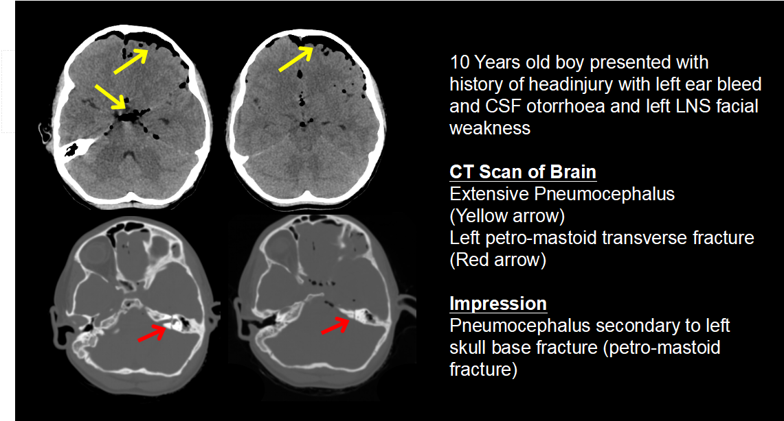
- Basilar skull fracture. This type of skull fractures involve a break in the bone at the base of the skull. These patients often have associated findings like, nerve injury, CSF leak, bruise on the face etc.
- Intracranial hematoma : Blood accumulated with the cranial cavity is often termed as intracranial hematoma. These can range from mild head injuries to life-threatening injuries. The different types of ICH are
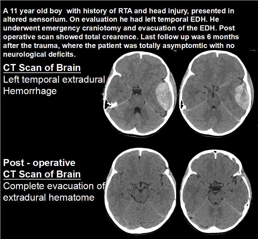
- Extradural hematoma: Blood collected underneath the skull, but on top of the dura(covering of the brain). They are usually associated with skull fracture. They are commonly seen in young adults and require emergency surgery if they are causing mass effect on the brain.
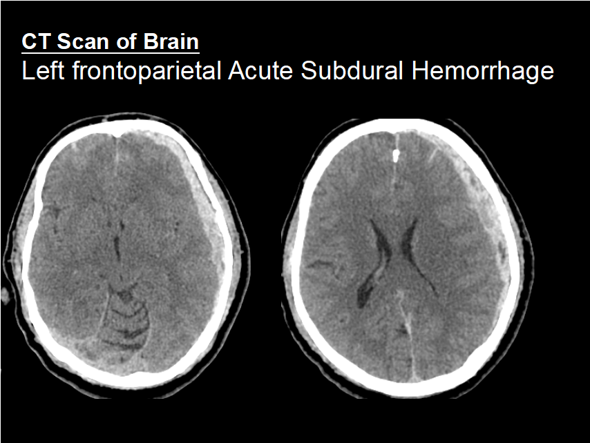
- Subdural hematoma: Blood collected underneath the dura, but outside of the brain. They are often associated with injury to the brain parenchyma. They require surgery if there are pressure effects on the brain.
- Contusion or intracerebral hematoma: A contusion is a bruise to the brain caused due to the brain hitting against the bony skull from within. Contusions may occur with skull fractures or other types of hematomas. Bleeding that occurs inside the brain parenchyma is calle3d intracerebral hematoma.
- Subarachnoid hematoma: Blood is collected between the brain parenchyma and the immediate layer covering the it (arachnoid).
- Diffuse axonal injury (DAI): They are usually caused by shaking of the brain within the cavity of the skull. Diffuse injuries can be mild, similar to a concussion, or may be very severe, where the patient is comatose. In DAI, the patient is usually in coma for a prolonged period of time, with injury to many different parts of the brain.
Diagnostic tests may include:
- Blood tests
- X-ray: A diagnostic test that uses X ray beams to produce images of internal tissues, bones, and organs.
- Computed tomography scan (CT scan): A procedure using X-rays and computer technology to produce 3 dimensional images of the body. They are good to show changes in the bone than changes in the soft tissue.
- Electroencephalogram (EEG): A procedure that records the brain’s electrical activity by means of electrodes placed onto the scalp.
- Magnetic resonance imaging (MRI): A diagnostic procedure that uses electromagnetic waves to produce detailed images of organs and structures within the body.
Treatment of Head Injury
Medical Management:
Aniconvulsants – Phenytoin, Levatricitam, Valproate, Clobazam, Lacosamideare among the commonly used anticonvulsants.
Antioedema – Mannitol, Lasix and hypertonic saline.
Cerebroprotective drugs – Cerebrolysin, citicholine, piracetam, etc
Other supportive medications – Analgesics, antacids, multivitamins etc
Observation: Injuries like cerebral concussion and small bleeding can be observed by just continuing medicines. GCS monitoring, neurological observation and sometimes ICP monitoring will be required.
Surgery: When there is significant mass effect and midline shift along with progressive worsening in the neurological state of the patient, the patient will need surgery.
Depressed fracture – Elevation and repair of depressed fracture
Extradural hematoma – Craniotomy and evacuation of EDH
Subdural Hematoma – Decompressive craniectomy and evacuation of subdural hematoma
Intracerebral hematoma – Craniotomy and evacuation of intracerebral hematoma, with or without decompressive craniectomy
Contusion – Craniotomy and evacuation of contusion, with or without decompressive craniectomy
Cranioplasty – the patients who have underwent decompressive craniectomy will need another surgery at a later date for replacement of the removed bone which is called cranioplasty.
Bone cranioplasty – replacing the patient’s own bone
Mesh cranioplsty – covering the brain surface with titanium moulded mesh or with custom made titanium mesh (3D printing).
GENERAL ENQURIES
186 0208 0208
+91 8884415615 | +91 8884458890
- gouthamcugati@gmail.com
- Narayana Multispecialty Hospital.
CAH/1, 3rd Phase, Devanur, Ring Road,
Mysore – 570019
OFFICE HOURS
| Monday – Saturday | 09:00 AM – 5:00 PM |