Cerebrovascular pathologies
STROKE
Stroke is a result of abrupt interruption of blood flow to the brain resulting in loss/disruption of neurological function. The interruption of blood flow can be caused by a blockage, leading to the more common ischemic stroke, or by bleeding in the brain, leading to the hemorrhagic stroke.
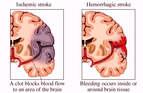
Ischemic stroke:
Ischemic stroke is by far the most common type of stroke, accounting for a large majority of strokes. The common blood vessel which gets blocked is the middle cerebral artery.
Hemorrhagic stroke
Symptoms
- Weakness or paralysis in your limbs, which may be on one or both sides
- Drooping of one side of face
- Dizziness and vertigo
- Confusion
- Vision problems, such as blindness in one eye or double vision
- Loss of coordination
Evaluation
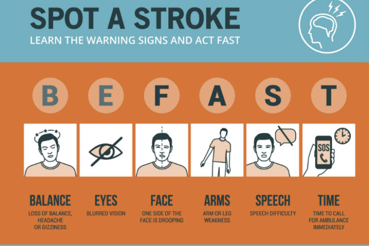
If someone is suspected to have stroke then do a quick evaluation by FAST
- Face. Is one side of their face drooping and difficult to move?
- Arms. Significant difficulty in raising either arm?
- Speech. Is their speech slurred or otherwise different?
- Time. If the answer to any of these questions is yes, it’s time to seek emergency medical service.
Intrinsic risk factors for Stroke include:
- High blood pressure
- Atherosclerosis
- High cholesterol
- Atrial fibrillation
- Previous heart attack
- Clotting disorders
- Congenital heart diseases
- Obesity
Extrinsic risk factors include:
- Diabetes
- Smoking
- Heavy alcohol misuse
- Use of certain drugs, such as cocaine
Diagnosis:
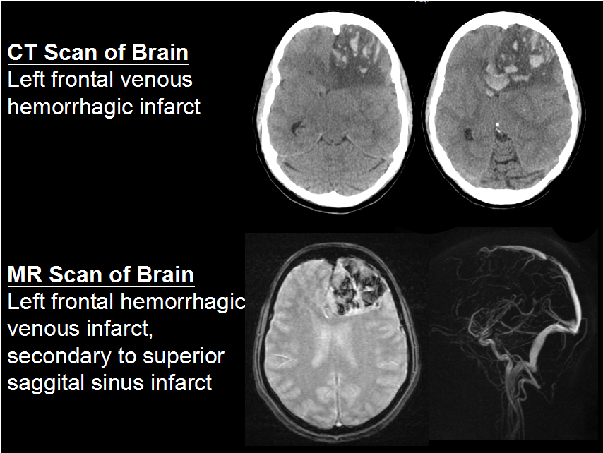
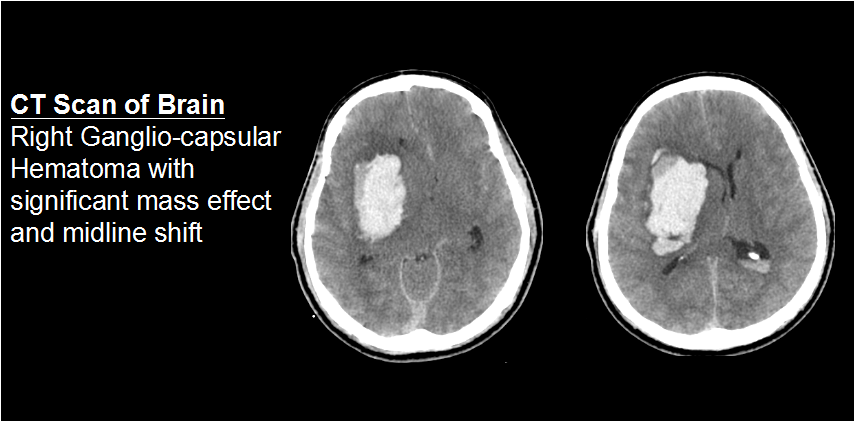
CT scan is ideal if hemorrhagic stroke is suspected
MRI of the brain is ideal if ischemic stroke is suspected
Treatment
Ischemic Stroke:
If the patient presents with in window period (3 hours) – tPA is recommended to reverse the stroke.
The other medications used are antiplatelet drugs, statins, cerebroprotective drugs and antti oedema drugs
Surgery – If there is severe swelling of the brain, then as a lifesaving procedure – Decompressive craniectomy is done.
Hemorrhagic stroke:
Medicines to reduce the high blood pressure. Anti-oedema and Cerebroprotecive drugs.
Surgery – If there is severe swelling of the brain or the volume of the bleeding is more then as a lifesaving procedure – Hematoma evacuation and Decompressive craniectomy is done.
Cerebral aneurysms
A cerebral aneurysm is a disorder where a blood vessel in the brain weakens, resulting in a bulging or ballooning of part of the vessel wall. The wall of the Aneurysm is thin and defective and is vulnerable for rupture, resulting in a catastrophic intracranial bleed.
The disorder may result from congenital defects or from other conditions such as high blood pressure, atherosclerosis (the buildup of fatty deposits in the arteries) or head trauma.

Symptoms
Unruptured aneurysms usually do not have any symptoms.
Ruptured aneurysms can present with many symptoms like headache, nausea, vomiting, stiff neck, double vision, photophobia, or loss of sensation. In more serious cases, the bleeding may cause brain damage, paralysis or coma.
Diagnosis
Cerebral aneurysms are identified by imaging modalities like MR angiogram, CT angiogram and Conventional angiogram.
Treatment
Surgery – Clipping of Aneurysm: An operation to “clip” the aneurysm is performed by doing a craniotomy (opening the skull surgically), and identifying the aneurysm and then isolating it from the bloodstream using one or more clips, which allows it to deflate.

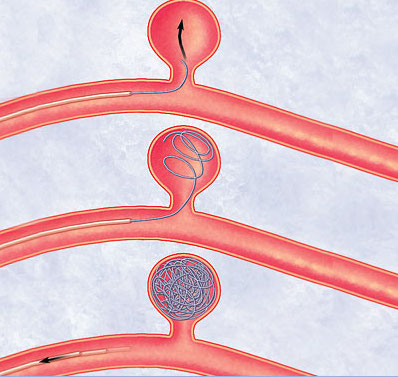
Endovascular coiling: A less invasive technique which does not require an operation, which uses micro catheters to deliver coils to the site of aneurysm from inside the blood vessel. Aneurysms may be treated with endovascular techniques when the risk of surgery is too high.
VASCULAR MALFORMATIONS
Vascular malformations are abnormal connection of an artery, vein or both. This is a result of abnormal development of blood vessels during the embryonal stage.
A typical malformation has abnormally dilated arteries leading directly to veins which a tortious and dilated.
There are different types of vascular malformations:
- Arteriovenous Malformation
- Cavernous Malformations
- Mixed Malformations
- Telangiectasis
- Vein of Galen Malformation
- Venous Malformations
Arteriovenous malformations
Vascular malformations of the brain may cause headaches, seizures, strokes, or bleeding in the brain. These blood vessels are enlarged, twisted, and tangled. Arteries and veins may be connected directly instead of being connected through fine capillaries.
Magnetic resonance Angiogram, computed tomography angiogram, conventional angiography can demonstrate vascular malformations.
Treatment options vary according to the severity and location of the malformation.
- Surgical removal
- Embolization (an operation in which pellets are used to block blood flow to and/or from the abnormal blood vessels)
- Irradiation – stereotactic radiosurgery or GKRS
- In some cases, treatment may not be necessary, just observation.
Cavernous malformations
Cavernous malformations or cavernomas present as abnormally enlarged collections of blood-filled spaces within the brain substance. A cavernous hemangioma acts like a “blood sponge” soaking up blood in-between capillaries, in the spaces between tissues larger cavernous spaces. Most of the times no treatment is required. If the patient has seizures, then one requires, antiepileptic medications. Surgery is required if intractable seizures or significant mass effect with neurological deficits.
GENERAL ENQURIES
186 0208 0208
+91 8884415615 | +91 8884458890
- gouthamcugati@gmail.com
- Narayana Multispecialty Hospital.
CAH/1, 3rd Phase, Devanur, Ring Road,
Mysore – 570019
OFFICE HOURS
| Monday – Saturday | 09:00 AM – 5:00 PM |
