Anatomy of the brain
The brain is a very important organ that controls every organ in the body. It can be called the CPU (central processing unit) of the human body which controls emotion, thought, memory, touch, temperature, motor skills, vision, breathing, sex, hunger, growth and every process that regulates our body.
The brain can be divided into the cerebrum, brainstem, and cerebellum:
- Cerebrum:The cerebrum (front of brain) comprises of the majority of the brain. It is composed of the right and left hemispheres, which are joined by the corpus callosum. The outermost part is called cerebral cortex and it controls initiation of movement, control of temperature, touch, smell, vision, hearing, judgment, reasoning, problem solving, emotions, and learning.
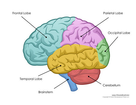
- Frontal lobe: The largest section of the cerebrum which is located in the front of the head, the frontal lobe is involved in movement. It also controls and defines personality, characteristics, smell and behaviour.
- Parietal lobe: The middle part of the brain, the parietal lobe helps a person to identify objects and understand geographical and spatial relationships. The parietal lobe is also involved in interpreting sensations like pain, touch, pressure and temperature in the body.
- Occipital lobe: The occipital lobe is the back part of the brain that is involved with interpreting vision.
- Temporal lobe: The temporal lobes form the sides of the brain, are involved in short-term memory, speech, musical rhythm, and some degree of smell recognition.
- Brainstem: The brainstem (middle of the brain) includes the midbrain, the pons, and the medulla. It is the part of the brain which has many vital centres and relay points. Functions of this area include: consciousness, movement of the eyes and mouth, relaying sensory signals, respirations, cardiac function, involuntary muscle movements, sneezing, coughing, vomiting, and swallowing.
- Midbrain: The topmost part of brainstem, which receives the fibres from the cerebrum tapering into the brain stem.
- Pons: The middle part of the brainstem, the pons has many control areas from the eye and face movements.
- Medulla: The lowest part of the brainstem and contains important control centres for the heartbeat and respiration.
- Cerebellum: The cerebellum is located at the back of the head. Below the cerebrum and behind the brain stem. It helps to coordinate voluntary muscle movements and to maintain posture, balance, and equilibrium.
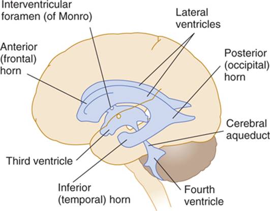
Ventricular System
The brain has fluid-filled cavities called ventricles. Inside the ventricles there are structures choroid plexus that produce clear colourless fluid called cerebrospinal fluid (CSF). CSF flows within and around the brain and spinal cord to lubricate it and help cushion it. This fluid is constantly produced and absorbed.
There are two ventricles deep within each cerebral hemispheres called the lateral ventricles. They both are connected with the third ventricle through an opening called the foramen of Monro. The third ventricle connects with the fourth ventricle through a narrow channel called the aqueduct of Sylvius. The fourth ventricle is located with the cerebellum. From the fourth ventricle, CSF flows into the subarachnoid space where it bathes the brain. CSF is absorbed into circulation by special filters called arachnoid villi.
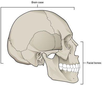
Skull
The skull is the bony protective structure around the brain. The skull is formed by fusion of eight bones. These bones include the two parietal, two temporal, one each of frontal, sphenoid, occipital and ethmoid.
Cranial nerves
There are twelve nerve which come out of the brain directly and carries signals and information. Rest of the information is transmitted through spinal cord.
- Olfactory nerve
- Optic nerve
- Occulomotor nerve
- Trocheal nerve
- Trigeminal nerve
- Abducent nerve
- Facial nerve
- Vestibulo-cochlear nerve
- Glossopharyngeal nerve
- Vagus nerve
- Accessory nerve
- Hypoglossal nerve
Meninges
The brain and spinal cord are covered by three protective layers of tissue called meninges. They are
- Outer dura mater
- Middle arachnoid mater
- Inner pia mater.
GENERAL ENQURIES
186 0208 0208
+91 8884415615 | +91 8884458890
- gouthamcugati@gmail.com
- Narayana Multispecialty Hospital.
CAH/1, 3rd Phase, Devanur, Ring Road,
Mysore – 570019
OFFICE HOURS
| Monday – Saturday | 09:00 AM – 5:00 PM |